Microvesicles (MVs), previously named microparticles, are small membrane enclosed structures released by activated and apoptotic cells by cell-surface membrane blebbing. Their size range from 100 to 1000 nm in diameter.
Microvesicles (MVs) lack a nucleus and contain a membrane skeleton. Once released, microvesicles (MVs) circulate in a variety of body fluids and vectorize information to multiple targets in the body.
MVs are diverse depending on their biogenesis and parent cell.
They are considered both as molecular signature of the cells from which they originate and as a depository of important biological information which can be exchanged with neighboring cells.
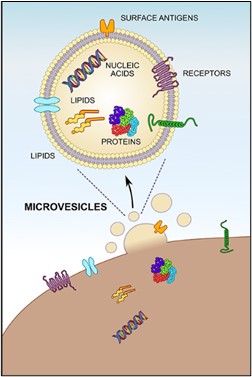
Circulating microvesicles are principally of platelet origin, but also derive from red blood cells, white blood cells and endothelial cells. Microvesicles express antigens originating from their parental cells on the surface, allowing for determination of the cell origin by the use of specifically labeled antibodies.
MVs have been described as procoagulant entities since their first report by Perter Wolf in 1967. This procoagulant activity relies to the presence of negative procoagulant phospholipids, mainly phosphatidylserine (PS), they can carry at their surface, on the external leaflet of the membrane.
In addition, the presence of Tissue Factor (TF) on subsets of MVs also significantly contributes to their procoagulant activity. A surface area unit of platelet MVs has approximately 50-to-100-fold higher procoagulant properties vs. an identical surface area unit of an activated platelet. Thus, the role of microvesicles in coagulation activation is well accepted. Overproduction of MVs has been related to various physiological and pathophysiological conditions.
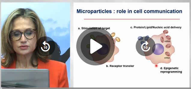
MVs are playing a role in blood coagulation, inflammation, cell activation and cancer metastasis.
Learn more about Microvesicles and watch our Webinar:
Microparticles: an emerging biomarker with multifaceted clinical indication
Download the Microvesicles Line brochure